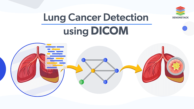
Overview and Steps for Lung Cancer Detection on DICOM Dataset
To train a machine learning model that can detect lung cancer from DICOM images.Steps of the Process
- Collection of Images in DICOM Format
- Conversion of the images and Labeling the Images
- Annotate all the Images
- Image pre-processing
- Image Augmentation
- Dividing the train and test data set
- Training of the Model
- Testing the Model
Solution Offered for Building the Detection Platform
The procedure starts from collecting the Images on which the model should be trained, then area which should be detected is label for the training set after that the annotation of the images are required which will be followed by pre-processing them. If the number of images are lesser than the required number of images than we can increase the number by image augmentation process. Next, the dataset will be divided into training and testing. Training the model will be done. The model can be ML/DL model but according to the aim DL model will be preferred. The model will be tested in the under testing phase which will be used to detect the detect the lung cancer the uploaded images.Machine Learning and Deep Learning Models
- Region-based Convolutional Network (R-CNN)
- Fast Region-based Convolutional Network (Fast R-CNN)
- Faster Region-based Convolutional Network (Faster R-CNN)
- Region-based Fully Convolutional Network (R-FCN)
- Neural Architecture Search Net (NASNet)
- Generative Adversarial Networks (GANs)
Architecture for Building Machine Learning Model for Cancer Detection
Steps for Building the Machine Learning Model based platform for Cancer Detection -- The first step to be followed is import new Images
- Next step is to label the images by using Labelme tool.
- Image Pre-processing
- Image Augmentation
- Dividing the train and test dataset
- Model Building (GAN)
- Training and Evaluation of GAN model


